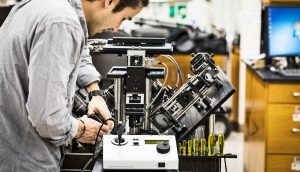
As part of their breast cancer treatment, women are subjected to lymph node dissection procedure. Doctors try to remove all cancerous tissues while protecting healthy breast tissue during this time. However, at present, there is no reliable approach to conclude whether the tissue excised is entirely cancer free at its edges. This acts as evidence that doctors have excised the entire tumor. Pathologists may require numerous days to process and examine the tissue with the use of conventional methods.
Owing to this, around 20–40% women have to undergo other subsequent breast-conserving surgeries to eliminate cancerous cells that were skipped during the primary procedure. A team of pathologists and mechanical engineers at the University of Washington has invented a new microscope that can be helpful in solving this as well as other issues. It can non-destructively and rapidly image the edges of big fresh tissue samples, in merely 30 Minutes, with the similar detail level as traditional pathology.
The new light-sheet microscope provides other benefits over the prevailing microscope technologies and processes. It saves valuable tissue for diagnosis and genetic testing, precisely and rapidly images the asymmetrical margins of large clinical samples, and enables pathologists to magnify and observe biopsy samples in 3D. The existing pathology methods include staining and processing tissue samples, implanting them in wax blocks, thin slicing, placing them on slides, then staining, and finally observing these 2D tissue sections with conventional microscopes—a procedure that can take days to produce results.
By contrast, the new microscope utilizes a light sheet optically “slice” through and images a tissue specimen without damaging any of it. The entire tissue is preserved for prospective downstream molecular analysis, which can produce additional useful data about cancer and result in more effective therapy decisions. The microscope can image surfaces of big tissue at high resolution and sew together several 2D images every second to rapidly produce a 3D image of a biopsy or surgical sample. That supplementary information can someday enable pathologists to more consistently and accurately grade and diagnose tumors.
These improvements were attained by the team by configuring several optical technologies in new approaches and enhancing them for clinical usage. Their open-top configuration, which sets all of the optics beneath a glass plate, lets them image bigger tissue specimens than other microscopes. At present, the team is functioning to boost the optical clearing process that permits light to infiltrate biopsy specimens more easily.
The study is published in the journal Nature Biomedical Engineering.
